


LncRNA regulates the progression of Triple-negative breast cancer
2Department of Oncology, the Rizhao People’s Hospital, Rizhao 276800, China
3Central Laboratory, the Rizhao People’s Hospital, Rizhao 276800, China
Abstract
1. Introduction
Triple
negative breast cancer (TNBC) is considered to be a heterogeneous and complex
subtype of breast cancer, with strong aggressive biological behavior, high
recurrence rate, high risk of distant metastasis, and poor prognosis. So far,
there is still the main focus of
public health attention. Managing TNBC in a clinical setting is difficult
because of its diverse nature, aggressive biological characteristics, and
absence of targeted treatments, leading to limited effectiveness of endocrine and
targeted therapies. The combination of surgery, radiation therapy, and
cytotoxic chemotherapy is the preferred method for treating TNBC patients. In
addition, resistance to traditional therapies may be caused by several
treatment cycles or intrinsic factors composed of cancer. Hence, individuals
are eagerly anticipating gaining a deeper insight into the patterns and
mechanisms of tumor development in TNBC cells, as well as discovering reliable
biomarkers for diagnosing and predicting outcomes for TNBC patients.
Long
non-coding RNAs (lncRNAs) are a
subclass of ncRNAs, consisting of over 200 nucleotides. The number
of LncRNAs greatly exceeds the number of protein coding genes in the human
genome. Reports indicate that lncRNAs are
crucial in the complex process of TNBC tumor formation and the development of
TNBC. The importance of lncRNAs in signaling specific cellular states,
detecting cellular pathologies like cancer, and guiding treatment decisions for
cancer patients is now widely acknowledged. LncRNA controls the aggressive
actions of TNBC cells through multiple intricate pathways, such as competitive
endogenous RNA (ceRNA) where lncRNA
competes with target genes to attach to miRNA, which is the primary mechanism
for many of the previously mentioned lncRNAs.
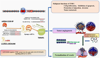
Our prior
studies have shown that lncRNA LINC01096 has the ability to interact with
miR-3130-3p [1],
leading to the derepression of TNFAIP1 by miR-3130-3p, thereby enhancing the
growth, infiltration, and movement of TNBC cells (Fig 1).
LINC01096 is abnormally expressed in TNBC, and LINC01096
can serve as a potential sponge ceRNA to adsorb miR-3130-3p. The miR-3130-3p
has the ability to attach to the 3'UTR of TNFAIP1 mRNA, leading to the
regulation of TNFAIP1 transcription by miR-3130-3p. LINC01096 is highly
expressed, while miR-3130-3p is lowly expressed. LINC01096 and
miR-3130-3p are associated with malignant biological
behaviors such as TNBC cell proliferation, invasion,
apoptosis inhibition, tumor angiogenesis and metastasis.
miR-3130-3p is down regulated and negatively regulated by LINC01096. In turn,
overexpression of miR-3130-3p can inhibit the growth of TNBC cancer cells and
tumor angiogenesis[2].
miR-3130-3p is expected to become a new target for breast cancer.
Identification of miR-3130-3p target interaction network has theoretical and
clinical significance. Following phosphorylation, it moves from the cytoplasm
to the nucleus where it is involved in critical processes including hypoxia
response, angiogenesis, DNA replication, and damage repair. It has the
characteristics of molecular changes and abnormal signaling pathways. It has
the characteristics of molecular changes and abnormal signaling pathways.
In recent years, research on lncRNAs in cancer has
significantly grown, making it a prominent subject in the field of RNA biology[3]. Because
of the tissue specific expression and up-regulated expression of lncRNA in
cancer, lncRNA represents an attractive target and biomarker for breast cancer
prognosis, therapeutic response, disease recurrence, and differentiation
between tumor and non tumor tissues. In addition, different subtype specific
lncRNAs can assist in diagnosis and subsequent treatment pathways, helping
patients stratify[4].
Fig. (1). LncRNA regulates the progression of TNBC. LINC01096 regulates
progression through miR-3130-3P/TNFAIP1 signaling pathway by sponge ceRNA by participating in cancer stemness, EMT, regulating cell
proliferation and apoptosis, invasion and metastasis, and participating in
angiogenesis regulation including tumor angiogenesis and vascular normalization
in tumor microenvironment hypoxia.
2. LncRNAs
involved in cancer stemness of TNBC
3. LncRNAs involved in TNBC EMT
4. LncRNAs
regulate cell proliferation and apoptosis
The
foundation of tumor growth lies in genetic mutations that cause cells to
multiply uncontrollably and prevent cell death[26]. Extensive research has also been conducted on lncRNAs involve TNBC cells proliferate and apoptosis. The primary focus of research is on
the ceRNA mechanism and network formation, which involves sequestering miRNA
and releasing its protein-coding counterpart from post-translational
regulation. This mechanism is primarily investigated as the key molecular
process underlying the biological functions of lncRNA[27]. Reports indicate that lncRNA GAS5 enhances TNBC
cell death and hinders growth through the regulation of miR-378a-5p/SUFU and miR-196a-5p signaling pathways[28–30]. In TNBC, HOST2 enhances cell proliferation and
migration through STAT3 by sequestering let-7b as a competitive endogenous RNA[31]. LINC00096 enhances cellular growth by binding to
miR-383-5p and controlling the levels of RBM3 in TNBC[32]. LINC00339/miR-377-3p/HOXC6 signaling pathway
promotes TNBC proliferation and inhibits cell apoptosis [33]. The lncRNA FAM83H-AS1 enhances the advancement of TNBC by controlling the miR-136-5p/metadherin pathway[34]. Furthermore, Multiple lncRNAs participate in regulation the
growth and cell death of TNBC cells, such as HCP5[35], LncRNA NRON[36,37], LUCAT1[38], HAND2-AS1[39–41], POU3F3[42]. Further research is needed to determine the
functional significance of these lncRNAs in TNBC cell proliferation and
apoptosis, making them potential candidates for future research. Cyclin dependent kinases (CDKs) control
the advancement of the cell cycle, being triggered by cyclin attachment and
blocked by CDK inhibitors[43,44]. TNBC is largely resistant to CDK4/6 inhibitors
due to the expression of CDK2/cyclin E, while free CDK2 inhibitors exhibit
normal tissue toxicity, limiting their therapeutic application in TNBC[44]. P27
inhibits CDK by binding to both the cyclin E/CDK2 complex and the cyclin
D/CDK4, 6 complex, playing a role in cell cycle regulation[45]. Reports indicate that LncRNA MIR100HG prevents G1
phase cell arrest by interacting with p27 to create RNA-DNA triple helix
structures. Within TNBC, it has been demonstrated that LUCAT can speed up the
cell cycle by controlling miR-5702[39]. Moreover, LncRNA RMST induces G0/G1
phase blockade in TNBC[46,47]. Additional mechanistic research is required to
explore how lncRNAs influence the advancement of the cell cycle in TNBC cells.
5. LncRNAs
regulate cell invasion and metastasis
6. LncRNAs
involved in angiogenesis
7. LncRNAs as a markers for the diagnosis of
TNBC
In order to investigate the role of lncrna in the
diagnosis of tnbc patients, many researchers have established study groups for
tnbc, with non tnbc patients and healthy individuals as control groups, to
study the differences in lncrna expression levels in tissue specimens, plasma,
and exosomes between different groups. It has now been discovered that there
are differential expressions of various lncrnas in tnbc compared to non tnbc
patients or healthy controls. For example, linc01096 is overexpressed
expression in tnbc, significantly higher than in normal breast tissue and
adjacent tissues, suggesting that linc01096 could be
useful as a marker for the diagnosis of tnbc[32]. Recently several studies also demonstrated that
there is increasing evidence shown lncrnas have diagnostic potential for
clinical tnbc patients. The expression levels of lncrnas x inactive-specific
transcript (xist) and nuclear paraspeckle assembly transcript 1 (neat1) were
significantly higher in the tnbc group than in the benign breast lesion and
healthy patients group[59], which could be used to distinguish tnbc from
other breast cancer types. Fan identified three lncrnas including ac091043.1,
ap000924.1 and foxcut maybe have strong diagnostic value for tnbc diagnosis by
comprehensive analysis of lncrna expression profiles and clinical data of 1097
breast cancer samples from tcga database[60]. The research results show that lncrna anril,
hif1a-as2, and uca1 have diagnostic value for tnbc, and the combined diagnostic
performance of the three is 0.934 in the area under the curve (auc), which is
better than the diagnostic effect of using them alone [61,62]
8. LncRNA
in prognostic evaluation of TNBC
8. Conclusion and future vision
References
1. Wang G-P, Mou Z-L, Xu Y-Y, Liu G-X, Wang
D-M, Zhang H-P. LINC01096 knockdown inhibits progression of triple-negative
breast cancer by increasing miR-3130-3p. Eur Rev Med Pharmacol Sci.
2019;23:7445–56.
2. Wang G-P, Mou Z-L, Xu Y-Y, Liu G-X, Wang
D-M, Zhang H-P. LINC01096 knockdown inhibits progression of triple-negative
breast cancer by increasing miR-3130-3p. Eur Rev Med Pharmacol Sci.
2019;23:7445–56.
3. Zhang W, Guan X, Tang J. The long
non-coding RNA landscape in triple-negative breast cancer. Cell Proliferation.
2021;54:e12966.
4. Brown JM, Wasson M-CD, Marcato P. The
Missing Lnc: The Potential of Targeting Triple-Negative Breast Cancer and
Cancer Stem Cells by Inhibiting Long Non-Coding RNAs. Cells. 2020;9:763.
5. Tu Z, Schmöllerl J, Cuiffo BG, Karnoub
AE. Microenvironmental Regulation of Long Noncoding RNA LINC01133 Promotes
Cancer Stem Cell-Like Phenotypic Traits in Triple-Negative Breast Cancers. Stem
Cells. 2019;37:1281–92.
6. Prabhu KS, Raza A, Karedath T, Raza SS,
Fathima H, Ahmed EI, et al. Non-Coding RNAs as Regulators and Markers for
Targeting of Breast Cancer and Cancer Stem Cells. Cancers (Basel). 2020;12:351.
7. Zeng Z, Fu M, Hu Y, Wei Y, Wei X,
Luo M. Regulation and signaling pathways in cancer stem cells: implications for
targeted therapy for cancer. Mol Cancer. 2023;22:172.
8. Jahangiri L, Ishola T, Pucci P,
Trigg RM, Pereira J, Williams JA, et al. The Role of Autophagy and lncRNAs in
the Maintenance of Cancer Stem Cells. Cancers (Basel). 2021;13:1239.
9. Osum M, Kalkan R. Cancer Stem Cells and
Their Therapeutic Usage. Adv Exp Med Biol. 2023;1436:69–85.
10. Sha S, Yuan D,
Liu Y, Han B, Zhong N. Targeting long non-coding RNA DANCR inhibits triple
negative breast cancer progression. Biol Open. 2017;6:1310–6.
11. Keshavarz M,
Asadi MH. Long non-coding RNA ES1 controls the proliferation of breast cancer
cells by regulating the Oct4/Sox2/miR-302 axis. FEBS J. 2019;286:2611–23.
12. Shin VY, Chen
J, Cheuk IW-Y, Siu M-T, Ho C-W, Wang X, et al. Long non-coding RNA NEAT1
confers oncogenic role in triple-negative breast cancer through modulating
chemoresistance and cancer stemness. Cell Death Dis. 2019;10:270.
13. Ma F, Liu X,
Zhou S, Li W, Liu C, Chadwick M, et al. Long non-coding RNA FGF13-AS1 inhibits
glycolysis and stemness properties of breast cancer cells through
FGF13-AS1/IGF2BPs/Myc feedback loop. Cancer Lett. 2019;450:63–75.
14. Alghazali MW,
Al-Hetty HRAK, Ali ZMM, Saleh MM, Suleiman AA, Jalil AT. Non-coding RNAs,
another side of immune regulation during triple-negative breast cancer. Pathol
Res Pract. 2022;239:154132.
15. Mittal V.
Epithelial Mesenchymal Transition in Tumor Metastasis. Annu Rev Pathol.
2018;13:395–412.
16. Zhang K, Liu
P, Tang H, Xie X, Kong Y, Song C, et al. AFAP1-AS1 Promotes
Epithelial-Mesenchymal Transition and Tumorigenesis Through Wnt/β-Catenin
Signaling Pathway in Triple-Negative Breast Cancer. Front Pharmacol.
2018;9:1248.
17. Li F, Xian D,
Huang J, Nie L, Xie T, Sun Q, et al. SP1-Induced Upregulation of LncRNA
AFAP1-AS1 Promotes Tumor Progression in Triple-Negative Breast Cancer by
Regulating mTOR Pathway. International Journal of Molecular Sciences.
2023;24:13401.
18. Youness RA,
Hafez HM, Khallaf E, Assal RA, Abdel Motaal A, Gad MZ. The long noncoding RNA
sONE represses triple-negative breast cancer aggressiveness through inducing
the expression of miR-34a, miR-15a, miR-16, and let-7a. J Cell Physiol.
2019;234:20286–97.
19. Youness RA,
Assal RA, Abdel Motaal A, Gad MZ. A novel role of sONE/NOS3/NO signaling
cascade in mediating hydrogen sulphide bilateral effects on triple negative
breast cancer progression. Nitric Oxide. 2018;80:12–23.
20. Fardi M,
Alivand M, Baradaran B, Farshdousti Hagh M, Solali S. The crucial role of ZEB2:
From development to epithelial-to-mesenchymal transition and cancer complexity.
J Cell Physiol. 2019;234:14783–99.
21. Zhang G, Li H,
Sun R, Li P, Yang Z, Liu Y, et al. Long non-coding RNA ZEB2-AS1 promotes the
proliferation, metastasis and epithelial mesenchymal transition in
triple-negative breast cancer by epigenetically activating ZEB2. J Cell Mol
Med. 2019;23:3271–9.
22. Tang J, Li Y,
Sang Y, Yu B, Lv D, Zhang W, et al. LncRNA PVT1 regulates triple-negative
breast cancer through KLF5/beta-catenin signaling. Oncogene. 2018;37:4723–34.
23. Wang L, Wang
R, Ye Z, Wang Y, Li X, Chen W, et al. PVT1 affects EMT and cell proliferation
and migration via regulating p21 in triple-negative breast cancer cells
cultured with mature adipogenic medium. Acta Biochim Biophys Sin (Shanghai).
2018;50:1211–8.
24. Nandagopal S,
Misra S, Sankanagoudar S, Banerjee M, Sharma P, Pane SE, et al. Long Non Coding
RNA in Triple Negative Breast Cancer: A Promising Biomarker in Tumorigenesis.
Asian Pac J Cancer Prev. 2023;24:49–59.
25. The Advanced
Research Progress of Tumor Immunotherapy. CCR [Internet]. 2024 [cited 2024 Jul
21]; Available from: http://www.cancercellresearch.org/v11n41.htm
26. Han G, Bai X,
Li F, Huang L, Hao Y, Li W, et al. Long non-coding RNA HANR modulates the
glucose metabolism of triple negative breast cancer via stabilizing hexokinase
2. Heliyon. 2024;10:e23827.
27. Wang Y, Bu N,
Luan X-F, Song Q-Q, Ma B-F, Hao W, et al. Harnessing the potential of long
non-coding RNAs in breast cancer: from etiology to treatment resistance and
clinical applications. Front Oncol. 2024;14:1337579.
28. Zheng S, Li M,
Miao K, Xu H. lncRNA GAS5-promoted apoptosis in triple-negative breast cancer
by targeting miR-378a-5p/SUFU signaling. J Cell Biochem. 2020;121:2225–35.
29. Li S, Zhou J,
Wang Z, Wang P, Gao X, Wang Y. Long noncoding RNA GAS5 suppresses triple
negative breast cancer progression through inhibition of proliferation and
invasion by competitively binding miR-196a-5p. Biomed Pharmacother.
2018;104:451–7.
30. Yan Y, Ma J,
Chen Q, Zhang T, Fan R, Du J. GAS5 regulated by FTO-mediated m6A modification
suppresses cell proliferation via the IGF2BP2/QKI axis in breast cancer. Discov
Oncol. 2024;15:182.
31. Hua K, Deng X,
Hu J, Ji C, Yu Y, Li J, et al. Long noncoding RNA HOST2, working as a
competitive endogenous RNA, promotes STAT3-mediated cell proliferation and
migration via decoying of let-7b in triple-negative breast cancer. J Exp Clin
Cancer Res. 2020;39:58.
32. Tian Y, Xia S,
Ma M, Zuo Y. LINC00096 Promotes the Proliferation and Invasion by Sponging
miR-383-5p and Regulating RBM3 Expression in Triple-Negative Breast Cancer.
Onco Targets Ther. 2019;12:10569–78.
33. Wang X, Chen
T, Zhang Y, Zhang N, Li C, Li Y, et al. Long noncoding RNA Linc00339 promotes
triple-negative breast cancer progression through miR-377-3p/HOXC6 signaling
pathway. J Cell Physiol. 2019;234:13303–17.
34. Han C, Fu Y,
Zeng N, Yin J, Li Q. LncRNA FAM83H-AS1 promotes triple-negative breast cancer
progression by regulating the miR-136-5p/metadherin axis. Aging (Albany NY).
2020;12:3594–616.
35. Wang L, Luan
T, Zhou S, Lin J, Yang Y, Liu W, et al. LncRNA HCP5 promotes triple negative
breast cancer progression as a ceRNA to regulate BIRC3 by sponging miR-219a-5p.
Cancer Med. 2019;8:4389–403.
36. Zhuo W, Lian
Z, Bai W, Chen Y, Xia H. 3D- and 2D-QSAR models’ study and molecular docking of
novel nitrogen-mustard compounds for osteosarcoma. Frontiers in Molecular
Biosciences [Internet]. 2023 [cited 2023 Jul 5];10. Available from:
https://www.frontiersin.org/articles/10.3389/fmolb.2023.1164349
37. Niu L, Fan Q,
Yan M, Wang L. LncRNA NRON down-regulates lncRNA snaR and inhibits cancer cell
proliferation in TNBC. Bioscience Reports [Internet]. 2019 [cited 2024 May
25];39. Available from: https://www.ncbi.nlm.nih.gov/pmc/articles/PMC6522729/
38. Mou E, Wang H.
LncRNA LUCAT1 facilitates tumorigenesis and metastasis of triple-negative
breast cancer through modulating miR-5702. Biosci Rep. 2019;39:BSR20190489.
39. Chen F, Zhang
Y, Chandrashekar DS, Varambally S, Creighton CJ. Global impact of somatic
structural variation on the cancer proteome. Nat Commun. 2023;14:5637.
40. McAleese CE,
Choudhury C, Butcher NJ, Minchin RF. Hypoxia-mediated drug resistance in breast
cancers. Cancer Letters. 2021;502:189–99.
41. Chandrashekar
DS, Karthikeyan SK, Korla PK, Patel H, Shovon AR, Athar M, et al. UALCAN: An
update to the integrated cancer data analysis platform. Neoplasia.
2022;25:18–27.
42. Yang J, Meng
X, Yu Y, Pan L, Zheng Q, Lin W. LncRNA POU3F3 promotes proliferation and
inhibits apoptosis of cancer cells in triple-negative breast cancer by
inactivating caspase 9. Biosci Biotechnol Biochem. 2019;83:1117–23.
43. Cheung A,
Chenoweth AM, Johansson A, Laddach R, Guppy N, Trendell J, et al. Anti-EGFR
antibody-drug conjugate carrying an inhibitor targeting CDK restricts
triple-negative breast cancer growth. Clin Cancer Res. 2024;
44. Sherr CJ,
Roberts JM. CDK inhibitors: positive and negative regulators of G1-phase
progression. Genes Dev. 1999;13:1501–12.
45. He W, Wang X,
Chen L, Guan X. A crosstalk imbalance between p27(Kip1) and its interacting
molecules enhances breast carcinogenesis. Cancer Biother Radiopharm.
2012;27:399–402.
46. Wang L, Liu D,
Wu X, Zeng Y, Li L, Hou Y, et al. Long non-coding RNA (LncRNA) RMST in
triple-negative breast cancer (TNBC): Expression analysis and biological roles
research. J Cell Physiol. 2018;233:6603–12.
47. Palcau AC,
Brandi R, Mehterov NH, Botti C, Blandino G, Pulito C. Exploiting Long
Non-Coding RNAs and Circular RNAs as Pharmacological Targets in Triple-Negative
Breast Cancer Treatment. Cancers (Basel). 2023;15:4181.
48. Jin X, Xu X-E,
Jiang Y-Z, Liu Y-R, Sun W, Guo Y-J, et al. The endogenous retrovirus-derived
long noncoding RNA TROJAN promotes triple-negative breast cancer progression
via ZMYND8 degradation. Sci Adv. 2019;5:eaat9820.
49. Qiao Y, Wang
B, Yan Y, Niu L. Long noncoding RNA ST8SIA6-AS1 promotes cell proliferation and
metastasis in triple-negative breast cancer by targeting miR-145-5p/CDCA3 to
inactivate the p53/p21 signaling pathway. Environ Toxicol. 2022;37:2398–411.
50. Qattan A.
Genomic Alterations Affecting Competitive Endogenous RNAs (ceRNAs) and
Regulatory Networks (ceRNETs) with Clinical Implications in Triple-Negative
Breast Cancer (TNBC). Int J Mol Sci. 2024;25:2624.
51. Han C, Li X,
Fan Q, Liu G, Yin J. CCAT1 promotes triple-negative breast cancer progression
by suppressing miR-218/ZFX signaling. Aging (Albany NY). 2019;11:4858–75.
52. Fu J, Dong G,
Shi H, Zhang J, Ning Z, Bao X, et al. LncRNA MIR503HG inhibits cell migration
and invasion via miR-103/OLFM4 axis in triple negative breast cancer. J Cell
Mol Med. 2019;23:4738–45.
53. Hu Q, Ye Y,
Chan L-C, Li Y, Liang K, Lin A, et al. Oncogenic lncRNA downregulates cancer
cell antigen presentation and intrinsic tumor suppression. Nat Immunol.
2019;20:835–51.
54. Zhang H, Zhang
N, Liu Y, Su P, Liang Y, Li Y, et al. Epigenetic Regulation of NAMPT by
NAMPT-AS Drives Metastatic Progression in Triple-Negative Breast Cancer. Cancer
Res. 2019;79:3347–59.
55. Ribatti D,
Nico B, Ruggieri S, Tamma R, Simone G, Mangia A. Angiogenesis and
Antiangiogenesis in Triple-Negative Breast cancer. Transl Oncol. 2016;9:453–7.
56. Wu S, Guo B, Zhang L, Zhu X, Zhao P,
Deng J, et al. A micropeptide XBP1SBM encoded by lncRNA promotes angiogenesis
and metastasis of TNBC via XBP1s pathway. Oncogene. 2022;41:2163–72.
57. Chen Y, Zhou Y, Chen J, Yang J, Yuan Y,
Wu W. Exosomal lncRNA SNHG12 promotes angiogenesis and breast cancer
progression. Breast Cancer. 2024;31:607–20.
58. Tao W, Sun W, Zhu H, Zhang J. Knockdown
of long non-coding RNA TP73-AS1 suppresses triple negative breast cancer cell
vasculogenic mimicry by targeting miR-490-3p/TWIST1 axis. Biochem Biophys Res
Commun. 2018;504:629–34.
59. Swellam M, El Magdoub HM, Shawki MA,
Adel M, Hefny MM, El-Shazly SS. Clinical impact of LncRNA XIST and LncRNA NEAT1
for diagnosis of high-risk group breast cancer patients. Curr Probl Cancer.
2021;45:100709.
60. Fan C-N, Ma L, Liu N. Comprehensive
analysis of novel three-long noncoding RNA signatures as a diagnostic and
prognostic biomarkers of human triple-negative breast cancer. J Cell Biochem.
2019;120:3185–96.
61. Liu M, Xing
L-Q, Liu Y-J. A three-long noncoding RNA signature as a diagnostic biomarker
for differentiating between triple-negative and non-triple-negative breast
cancers. Medicine (Baltimore). 2017;96:e6222.
62. Q Y, Y F, J W, H Y, X Z. Roles of lncRNA
in the diagnosis and prognosis of triple-negative breast cancer. Journal of
Zhejiang University Science B [Internet]. 2023 [cited 2024 Jul 19];24.
Available from: https://pubmed.ncbi.nlm.nih.gov/38057269/
63. Fan C-N, Ma L, Liu N. Comprehensive
analysis of novel three-long noncoding RNA signatures as a diagnostic and
prognostic biomarkers of human triple-negative breast cancer. J Cell Biochem.
2019;120:3185–96.
64. Zhu T, Wang J, Li J, Zhang Q, Shang Y,
Zhou J, et al. A serum LncRNA signature for predicting prognosis of
triple-negative breast cancer. Clin Chim Acta. 2023;549:117535.
65. Zhang S, Ma F, Xie X, Shen Y. Prognostic
value of long non-coding RNAs in triple negative breast cancer: A
PRISMA-compliant meta-analysis. Medicine (Baltimore). 2020;99:e21861.
Share and Cite
Ren Z. J., Zeng D.R., Wang P. Q., Wang R., Zhou B. Q., Zhang Y. The identification of potential inhibitors of SARS-CoV-2 spike protein by virtual screening FDA-approved compound library. Chronic Diseases Prevention Review 2024, 8 (29), 1. DOI: 10.54762/CDPR2024.29.1-4.
Ren Z. J., Zeng D.R., Wang P. Q., Wang R., Zhou B. Q., Zhang Y. (2024). The identification of potential inhibitors of SARS-CoV-2 spike protein by virtual screening FDA-approved compound library. Chronic Diseases Prevention Review, 8(29), 1. https://doi.org/10.54762/CDPR2024.29.1-4
Chicago/Turabian StyleRen Z. J., Zeng D.R., Wang P. Q., Wang R., Zhou B. Q., Zhang Y. 2024. "The identification of potential inhibitors of SARS-CoV-2 spike protein by virtual screening FDA-approved compound library." Chronic Diseases Prevention Review 8 (29):1. doi: 10.54762/CDPR2024.29.1-4.